When your pet faces a surgical procedure, the stakes could not be higher. As a loving pet owner, you want every possible advantage working in your favor before your companion goes under anesthesia. That is where advanced veterinary imaging becomes one of the most powerful tools available to modern veterinary medicine. For pet owners in Huntington, NY and across Long Island, access to state-of-the-art diagnostic imaging for pets is no longer a luxury reserved for specialty hospitals in major cities. It is a critical step that can mean the difference between a successful outcome and an avoidable complication.
Veterinarians today rely on a range of imaging technologies to see what cannot be detected during a physical exam alone. From digital X-rays to ultrasound, and more recently, veterinary CT scans and pet MRI and CT imaging, the diagnostic landscape has changed dramatically. Understanding why these tools matter before surgery can help you make more informed decisions as a pet owner and advocate for your animal’s health.
What Diagnostic Imaging Reveals That Physical Exams Cannot
A thorough physical examination is always the starting point of good veterinary care, but it has its limitations. The human hand and eye can only detect so much. Internal structures, soft tissue abnormalities, early-stage tumors, vascular anomalies, and subtle bone fractures often go undetected without imaging support.
Diagnostic imaging for pets on Long Island has expanded significantly in recent years, giving local veterinarians and specialists a clearer picture of what is happening inside an animal’s body before any surgical incision is made. When a dog presents with unexplained lameness, for example, a standard X-ray might reveal a bone abnormality. But a veterinary CT scan in Huntington, NY can show a three-dimensional rendering of the joint, surrounding tissue, and bone density in a way that flat imaging simply cannot replicate.
This level of detail matters enormously in surgical planning. A surgeon who enters a procedure with a precise, multi-dimensional understanding of the anatomy involved is far better prepared than one relying on generalized knowledge and two-dimensional images. Pre-surgical imaging helps identify the exact location of a mass, determine whether it has spread to neighboring tissue, assess the blood supply involved, and evaluate the structural integrity of bones and joints. Each of these data points directly influences how a surgeon plans the approach, what instruments to use, and what to expect once the procedure begins.
For cats, small animals, and exotic species, this level of pre-operative insight is especially valuable. These patients often cannot communicate discomfort clearly, and their smaller anatomy demands greater precision from the surgical team. Imaging removes guesswork and replaces it with knowledge.
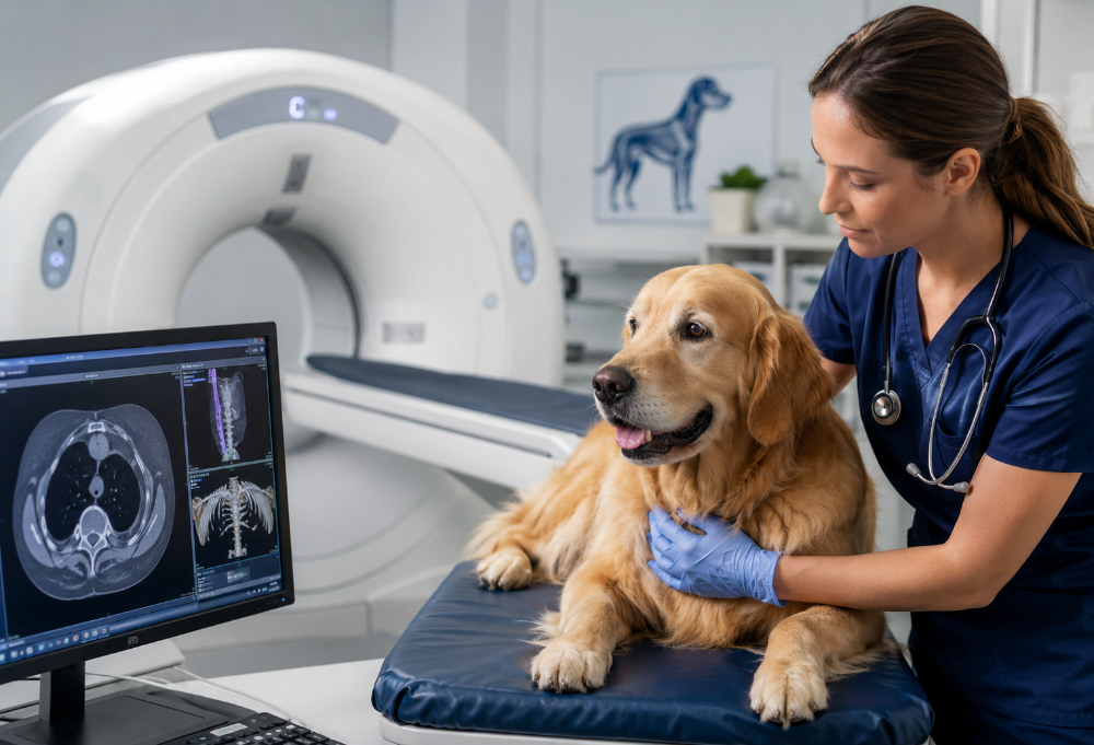
How Veterinary CT Scans Are Changing Pre-Surgical Planning
Among the imaging modalities now available in veterinary medicine, the CT scan has arguably transformed pre-surgical planning more than any other tool. A veterinary CT scan uses a series of X-ray images taken from multiple angles and processed by computer software to generate detailed cross-sectional views of the body. In Huntington, NY and surrounding areas of Long Island, veterinary practices offering this technology are helping surgeons prepare with a level of confidence that was simply not possible a generation ago.
CT imaging is particularly valuable for evaluating the thorax and abdomen, identifying foreign bodies, assessing trauma injuries, and staging cancer. When a pet is diagnosed with a tumor, for instance, a CT scan can reveal whether the mass is localized or has spread to the lymph nodes or distant organs. This information directly determines whether surgery is the right course of action at all, and if so, what the surgical goals should realistically be.
Orthopedic surgeries benefit enormously from CT-guided planning as well. Hip dysplasia corrections, spinal surgeries, and complex fracture repairs all require a thorough understanding of three-dimensional anatomy. Advanced veterinary imaging gives surgeons a virtual roadmap before they ever pick up a scalpel. In many cases, this has been shown to reduce surgical time, minimize trauma to surrounding tissue, and support faster recovery for the patient.
The process of undergoing a CT scan is also relatively straightforward for most pets. Short-duration anesthesia is typically required to keep the animal still during the procedure, but the scan itself is quick. The resulting data can be reviewed immediately and shared with specialists for consultation, which is particularly useful in complex cases that may benefit from a second expert opinion.
The Role of Pet MRI and CT Imaging in Neurological and Soft Tissue Cases
While CT scans excel at imaging bone and dense tissue, pet MRI and CT imaging together provide a more complete picture for cases involving the nervous system and soft tissue. Magnetic resonance imaging uses powerful magnets and radio waves to create detailed images of soft tissue structures, making it the gold standard for evaluating the brain, spinal cord, and intervertebral discs.
If a dog is showing signs of neurological dysfunction, such as sudden weakness in the hind limbs, seizures, or unexplained behavioral changes, an MRI can identify lesions, inflammation, herniated discs, or tumors that would not be visible on a CT scan alone. For pets on Long Island facing these kinds of diagnoses, having access to both MRI and CT imaging under one roof streamlines the diagnostic process and reduces the stress of transporting an already compromised animal to multiple facilities.
Combining both modalities when appropriate gives the surgical team a comprehensive view of the patient’s condition. A veterinarian might use a CT scan to evaluate bony involvement and an MRI to assess the degree of soft tissue or spinal cord compression. Together, these images inform a surgical plan that is precise, targeted, and grounded in real anatomical data rather than assumptions.
Soft tissue surgeries involving the liver, kidneys, bladder, or reproductive organs also benefit from pre-surgical MRI evaluation. The ability to delineate tumor margins, identify vascular involvement, and assess organ function before opening a patient is an enormous advantage. It allows surgeons to have more honest, informed conversations with pet owners about likely outcomes and potential risks, which is essential for trust and shared decision-making.
Choosing a Veterinary Facility With Advanced Imaging Capabilities
Not all veterinary practices offer the same level of diagnostic technology, and that gap matters when your pet needs surgery. Pet owners in Huntington, NY and throughout Long Island should ask prospective veterinarians and surgical teams specific questions about what imaging technologies are available on-site and how pre-surgical imaging is integrated into the standard of care.
Facilities that invest in advanced veterinary imaging demonstrate a commitment to evidence-based medicine and patient safety. They are not simply offering convenience; they are raising the standard of care for every patient who walks through the door. When evaluating a surgical team, consider whether they use diagnostic imaging for pets routinely as part of their pre-operative protocol, or only in exceptional cases. The former approach reflects a proactive philosophy that prioritizes preparation and precision.
Ask whether the imaging is interpreted by a board-certified radiologist, either on-site or through a teleradiology service. The quality of the image is only as valuable as the quality of the interpretation. Facilities that partner with veterinary radiologists to review CT and MRI findings bring an additional layer of expertise to the diagnostic process that can catch details a generalist might miss.
Cost is often a concern for pet owners, and it is a fair one to raise. Advanced imaging does add to the overall cost of a surgical workup. However, the cost of a preventable complication, an unexpected finding discovered mid-surgery, or a procedure that could have been avoided with better pre-operative information is almost always greater. Investing in comprehensive pre-surgical imaging is, in most cases, an investment in a better outcome.
Conclusion
For pet owners in Huntington, NY and across Long Island, advanced veterinary imaging is one of the most meaningful steps you can take before your pet undergoes surgery. From veterinary CT scans to pet MRI and CT imaging, these tools give surgical teams the knowledge they need to operate with confidence and care. When you choose a practice that prioritizes diagnostic imaging for pets, you are choosing a higher standard of medicine for the animal who depends on you most.
Need Veterinary Surgical Care Near You?
At Veterinary Surgical Center of Long Island, we understand how important your pet’s health is, which is why we are here to provide expert care during emergencies and complex surgical procedures. Whether your pet is facing a critical emergency or needs specialized surgery, our team in Huntington, NY, is ready to offer life-saving treatment with the latest technology and compassionate care. If your pet requires emergency attention or advanced surgical care, don’t hesitate to reach out to us. We’re committed to working alongside you and your primary care veterinarian to create a personalized treatment plan that ensures the best possible outcome for your furry family member. Contact us today and let us help your pet get back to a healthier, happier life.